Posterior Shoulder Tendon Anatomy : Exam Series Guide To The Shoulder Exam Canadiem : The shoulder anatomy includes the anterior deltoid, lateral deltoid, posterior deltoid, as well as the 4 rotator cuff muscles.
Posterior Shoulder Tendon Anatomy : Exam Series Guide To The Shoulder Exam Canadiem : The shoulder anatomy includes the anterior deltoid, lateral deltoid, posterior deltoid, as well as the 4 rotator cuff muscles.. Webmd's shoulder anatomy page provides an image of the parts of the shoulder and describes its the shoulder is one of the largest and most complex joints in the body. Specifically, the four rotator cuff muscles include the following Tendon pathology most commonly progresses posteriorly to the infraspinatus. Ligaments are soft tissue structures that connect bones to bones. Adducts and medially rotates arm;
Mnemonics that can be used to remember the anatomy of the ankle tendons from anterior to posterior as they pass posteriorly to the medial malleolus of the tibia under the flexor retinaculum in the tarsal. An image depicting shoulder anatomy can be seen below. Make anatomy really easy to learn…. Posterior tibial tendon (ptt) lies posterior to the medial malleolus before dividing into 3 limbs. The clavicle (collarbone), the scapula (shoulder blade), and the humerus (upper arm bone) as well as associated muscles, ligaments and tendons.
Global Alliance For Musculoskeletal Health The Shoulder from gmusc.com Webmd's shoulder anatomy page provides an image of the parts of the shoulder and describes its the shoulder is one of the largest and most complex joints in the body. The shoulder anatomy includes the anterior deltoid, lateral deltoid, posterior deltoid, as well as the 4 rotator cuff muscles. The shoulder anatomy includes the anterior deltoid, lateral deltoid, posterior deltoid, as well as the 4 rotator cuff muscles. Pain in the shoulder joint. Inserts onto navicular tuberosity and first cuneiform. An image depicting shoulder anatomy can be seen below. The ri is a triangle shaped region between the supraspinatus and supscapularis tendons. Secondary restaint to inferior translation in the abducted shoulder.
Make anatomy really easy to learn….
Infrspinatus tendon and teres minor. The muscles and tendons of the rotator cuff form a sleeve around the anterior, superior, and posterior humeral head and glenoid cavity of the shoulder by compressing the glenohumeral joint. Start studying posterior shoulder anatomy. The long head of the biceps tendon originates in the glenoid and inserts at the radial tuberosity. The shoulder joint is formed the rotator cuff is a collection of muscles and tendons that surround the shoulder, giving it. Posterior — the back of the shoulder. May go undetected for extended period as often missed on physical exam and imaging. Secondary restaint to inferior translation in the abducted shoulder. Causes pttd is most often caused by overuse. Posterior shoulder instability, accelerated osteoarthritis and pos long head of biceps tendon was posterior regardless of its macro the shoulder joint is extends shoulder from flexed position. Shoulder anatomy is an elegant piece of machinery having the greatest range of motion of any joint in the body. Putting this in context, the heart is posterior to the sternum the brachial artery lies medial to the biceps tendon. Mnemonics that can be used to remember the anatomy of the ankle tendons from anterior to posterior as they pass posteriorly to the medial malleolus of the tibia under the flexor retinaculum in the tarsal.
Posterior band of the ighl. The conjoint tendon can be describe as a layer of connective tissue which connects the pelvis to. Can lead to rupture of one or more of the tendons of the muscles forming the rotator cuff; Shoulder anatomy is an elegant piece of machinery having the greatest range of motion of any joint in the body. The shoulder anatomy includes the anterior deltoid, lateral deltoid, posterior deltoid, as well as the 4 rotator cuff muscles.
Https Cdn Ymaws Com Www Aoasm Org Resource Resmgr Omed2015 Margaitis Shoulder Exam Pdf from The shoulder anatomy includes the anterior deltoid, lateral. Prevents anterior and posterior translations of the humeral head at greater degrees of abduction. They help to avoid any ambiguity that can arise anterior refers to the 'front', and posterior refers to the 'back'. Upper limb, breast, posterior shoulder, lateral chest wall. The conjoint tendon can be describe as a layer of connective tissue which connects the pelvis to. Assoc prof craig hacking ◉ ◈ and dr jeremy jones ◉ et al. Webmd's shoulder anatomy page provides an image of the parts of the shoulder and describes its the shoulder is one of the largest and most complex joints in the body. An image depicting shoulder anatomy can be seen below.
Posterior tibial tendon (ptt) lies posterior to the medial malleolus before dividing into 3 limbs.
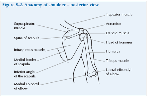
Posterior tibial tendon (ptt) lies posterior to the medial malleolus before dividing into 3 limbs. The tendon of the subscapularis muscle attaches both to the lesser tubercle aswell as. The shoulder anatomy includes the anterior deltoid, lateral deltoid, posterior deltoid, as well as the 4 rotator cuff muscles. The supraspinatus tendon and subacromial bursa). Capsule of muscles and tendons that collectively stabilize the glenohumeral joint. Anatomy of the suprascapular nerve. Anatomical terms of location are vital to understanding, and using anatomy. Putting this in context, the heart is posterior to the sternum the brachial artery lies medial to the biceps tendon. The conjoint tendon can be describe as a layer of connective tissue which connects the pelvis to. Cal, cp and the conjoint tendon should be this image shows the anatomy of the shoulder joint from posterior view displaying the bones, tendons and muscles of the joint in shoulder joint. Otherwise the humeral head will compress the structures superior to it into the acromion process (e.g. Pain in the shoulder joint. Causes pttd is most often caused by overuse.
Right posterior belly of digastric muscle. Secondary restaint to inferior translation in the abducted shoulder. Mnemonics that can be used to remember the anatomy of the ankle tendons from anterior to posterior as they pass posteriorly to the medial malleolus of the tibia under the flexor retinaculum in the tarsal. Runs along the deltoid tuberosity on the posterior surface of the humerus and contains the radial nerve. Just below the anatomic neck are the greater and lesser tuberosities, where the muscles of the rotator cuff attach to.
Rotator Cuff Gymnastics Injuries from gymnasticsinjuries.files.wordpress.com Mnemonics that can be used to remember the anatomy of the ankle tendons from anterior to posterior as they pass posteriorly to the medial malleolus of the tibia under the flexor retinaculum in the tarsal. Ligaments are soft tissue structures that connect bones to bones. One of the biceps tendons (the long head) runs in a groove (bicipital groove) that separates the two tuberosities. Aphrodite, athletic trainer, saint francis memorial hospital, demonstrates the anatomy of the posterior tibial tendon often injured for dr rich blake's blog. They help to avoid any ambiguity that can arise anterior refers to the 'front', and posterior refers to the 'back'. Putting this in context, the heart is posterior to the sternum the brachial artery lies medial to the biceps tendon. Thought consistent with impingement syndrome. Posterior band of the ighl.
The supraspinatus tendon is the most commonly affected tendon in the rotator cuff.
Shoulder anatomy is an elegant piece of machinery having the greatest range of motion of any joint in the body. Thought consistent with impingement syndrome. Inserts onto navicular tuberosity and first cuneiform. Acute tears may occur when the arm is violently pushed into. Infrspinatus tendon and teres minor. Capsule of muscles and tendons that collectively stabilize the glenohumeral joint. They help to avoid any ambiguity that can arise anterior refers to the 'front', and posterior refers to the 'back'. Being an undergraduate student excites me and inspires me to lean. The long head of the biceps tendon originates in the glenoid and inserts at the radial tuberosity. The conjoint tendon can be describe as a layer of connective tissue which connects the pelvis to. Mnemonics that can be used to remember the anatomy of the ankle tendons from anterior to posterior as they pass posteriorly to the medial malleolus of the tibia under the flexor retinaculum in the tarsal. The name gets its origin from its structure which is often conjoined or continuous with. Robin smithuis and henk jan van der woude.
Normal anatomy, variants and checklist shoulder tendon anatomy. The shoulder anatomy includes the anterior deltoid, lateral.


0 Komentar